In nuclear architecture, understanding how DNA is packed and how it is organized plays an important role in understanding the disease etiology, which typically requires millions of cells as a starting material to generate high-resolution 3D genome maps.
However, for Hutchinson-Gilford Progeria Syndrome (HGPS), a rare genetic condition, the primary patient-derived cells are fragile, slow-growing, and finite. This biological scarcity has restricted the use of Hi-C technology, the gold standard for 3D genome mapping, creating a knowledge gap regarding how the nuclear architecture is associated with this disease, specifically in the fat cell development process.
Recognizing this bottleneck, molecular biologist Nishita Gogia re-engineered the technology to suit the limited, scarce cell material available. Her work provides a methodological bridge between the limitations of rare disease samples and the data-intensive demands of modern genomics.
The Data Scarcity Problem in Rare Disease
Progeria is a premature/accelerated aging syndrome caused by a single point mutation in the LMNA gene, which produces the abnormal, truncated protein progerin. Progerin destabilizes the nuclear envelope, misshaping the nucleus and disorganizing the chromatin inside the nucleus.
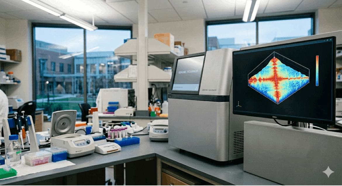
Researchers used High-throughput Chromosome Conformation Capture (Hi-C) to map the genome’s 3D spatial contacts. While the lab had expertise in Hi-C, applying it to primary patient-derived cells was new. Standard Hi-C needs five to ten million cells, typically from cell lines. However, progeria fibroblasts are “primary” cells with a short lifespan. Expanding them for standard Hi-C risks of losing the culture or cell death.
Engineering the Low-Input Workflow
Gogia engineered a low-input genomic workflow. This required a ground-up recalibration of the standard sequencing protocol. Her initial task was foundational: architecting optimized 2D culture protocols that allowed these delicate primary fibroblasts to grow efficiently for significantly longer periods than previously achievable.
“Working with primary patient-derived material is fundamentally different from working with established cell lines,” Gogia explains. “Everything is harder, the yields are lower, and there is almost zero margin for error. If the culture conditions aren’t perfect, you lose months of work before the experiment even begins.”
For weeks, Gogia tested varying media conditions, passage timings, and seeding densities. The goal was to keep the cells viable long enough to reach a critical mass, even if that mass was far below the industry standard for genomics. Once the culture protocols were stabilized, she turned her attention to the Hi-C workflow itself.
The standard protocol involves several high-loss steps: cell lysis, restricting digestion of the DNA, biotin labeling, and ligation. In a sample of five million cells, losing 20% of the material at each step is acceptable. In a sample of 50,000 cells, it is catastrophic. Gogia had to rethink the protocol at every stage to minimize loss and maximize “signal-to-noise” ratio.
Navigating Technical Obstacles
Gogia overcame significant technical hurdles to successfully apply Hi-C to low-input samples, particularly primary patient-derived cells. The first step of calibration was to isolate the nucleus from the cells, which required calibration, as it would significantly reduce the debris carried over from the whole cell mass, further standardizing the cross-linking conditions and tweaking reaction times across the Hi-C protocol led to a successful protocol execution of these primary cells. The final challenge was library preparation. Gogia noted the difficulty is preserving the genome’s architectural signal over technical noise, especially with minimal material, where errors are magnified. This landmark achievement allows researchers to study 3D genome organization in a disease-relevant context without cell immortalization, which can mask aging signatures.
Impact on the Regulatory and Scientific Landscape
The broader significance of this work is tied to the evolving landscape of genomic medicine, specifically as regulatory bodies and funding agencies like the National Institutes of Health (NIH) emphasize the transition toward “disease-relevant” models. While immortalized cell lines have served as the backbone of research for decades, they are often poor proxies for the actual physiological state of a patient. By bridging this gap, Gogia’s innovation aligns with a global shift toward precision medicine, where the focus is on the individual rather than the average.
Crucially, the low-input Hi-C protocol engineered by Gogia is not a localized success limited to a single laboratory; it is a replicable methodological blueprint with global implications. By providing a validated pathway to study the 3D genome in primary patient cells, she has created a toolset that can be exported to any research context where patient material is scarce. This standardized approach can now be utilized for other laminopathy models affecting cardiac and muscular health, rather than being restricted solely to HGPS. .
Within the organization, the impact of this new standard was immediate. The low-input workflow became a foundational tool that transformed the lab’s strategic capabilities, allowing the team to move to investigate how a specific patient’s mutation physically alters their genomic layout.
“When you can finally see the physical layout of the genome in a patient-derived cell, you’re not just looking at a list of genetic instructions,” Gogia reflects. “You’re seeing the structural foundation of life. If the foundation is misaligned, every downstream process, from how the cell reads its own genes to how it repairs its own damage, is compromised. By standardizing the way we see these failures, we aren’t just solving a problem for one lab; we’re giving the global research community a way to finally see the core of these diseases in high resolution.”
A Legacy of Methodological Innovation
Nishita Gogia’s role in this project was far more than that of a traditional graduate researcher. In an environment where most peers were executing established protocols, she was an innovator of the protocols themselves. Her ability to operate at the intersection of three challenging domains, primary cell biology, high-resolution genomics, and rare disease pathology, distinguishes her work from standard academic contributions.
Key outcomes include a stabilized research pipeline, a validated low-input technology platform, and new insights into the spatial biology of aging specifically in the context of fat cell formation. This research has been presented at prestigious venues like Cold Spring Harbor Laboratory and the Progeria Research Foundation, where the utility of her low-input approach was recognized.
Looking Forward: The Future of Genomic Mapping
As the scientific community moves toward a more nuanced understanding of “epigenomics”, the study of how the genome is regulated beyond just the DNA sequence, spatial organization has become a central pillar of research. The tools developed by Gogia ensure that researchers studying the rarest of diseases are not left behind in this genomic revolution.
The “Architecture of Aging” is no longer a metaphorical concept; thanks to the technical ingenuity of researchers like Gogia, it is a mapped reality. By engineering a solution to a physical limitation, she has ensured that the most vulnerable cells can finally tell their story in high resolution.
“The goal isn’t just to produce a single map,” Gogia says. “It’s to build a set of tools that can handle the complexity of human biology as it actually exists, scarce, fragile, and difficult to capture. If we can map the most challenging cells in the world, we can map anything.”
The institution continues to build on this foundation, utilizing the workflows Gogia established to investigate potential therapeutic targets that could one day “re-stabilize” the warped genomes of progeria patients. In the high-stakes world of rare disease research, where every cell counts, Gogia’s contributions have provided the field with a much-needed margin for success.